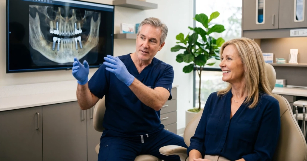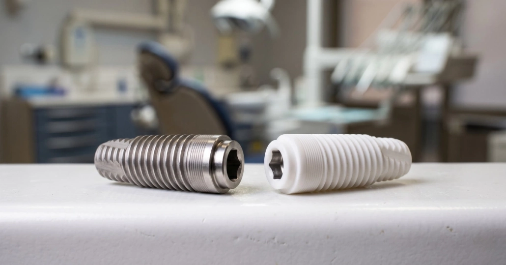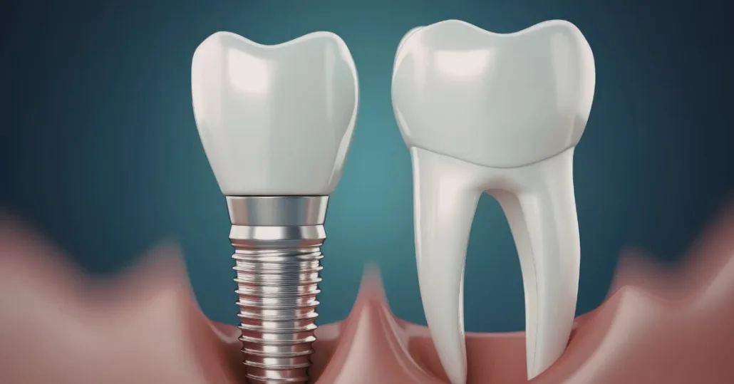
“Do I need a bone graft for dental implants” is one of the most common questions I hear in consultations, usually right after a patient has been told by another office that grafting is their only path forward. In many of those cases, it is not the full picture. I have placed enough zygomatic and pterygoid implants over the years to know that “you need a bone graft” is sometimes an incomplete answer rather than a final one. A cone beam CT scan tells us which is true for your jaw specifically, and that distinction can change your timeline, your cost, and sometimes your entire treatment plan.
| 25% Bone width that can be lost in the first year after tooth extraction |
3-6 Months of typical healing after a bone graft, before implant placement |
4-6 Implants typically used in a full-arch zygomatic or Stabili-Teeth® restoration |
What Actually Determines Whether You Need a Bone Graft for Dental Implants
Bone grafting for dental implants exists to solve one problem – not enough bone volume, height, or density at the exact site where an implant needs to sit. A standard X-ray cannot measure that with any real accuracy. A cone beam CT scan can, and it is the only reliable way to know whether grafting applies to your situation before a treatment plan is built around it.
What Your CT Scan Evaluates Before Any Grafting Decision
- Bone height at each potential implant site, measured against the inferior alveolar nerve (lower jaw) or the sinus floor (upper jaw)
- Bone width, since a site can have enough height but still lack the horizontal ridge a standard implant needs
- Bone density, graded on the Misch D1 through D4 scale
- Nerve and sinus position, which shapes where an implant can safely go
- Root remnants or old pathology that would not show up on a flat film
For a single missing tooth in a spot with real, localized bone loss, a graft is often genuinely the right call, and a fairly routine one. For patients missing most or all of their teeth across an arch, there is frequently another path that avoids grafting altogether. That path is covered further down.
How Bone Grafting Works, What It Costs, and How Long It Takes
Finding insufficient bone on a CT scan is not a dead end. It is the start of a staged plan, and bone grafting for implants is a well-established procedure with predictable healing, not an experimental one.
AutograftBone taken from another site in your own body, often the chin or the back of the jaw. Carries live cells that can support integration, at the cost of a second surgical site. |
AllograftProcessed donor bone from a bone bank, treated to preserve the mineral structure that encourages new bone growth. Widely used and well studied. |
XenograftBone material derived from bovine sources, with the organic components removed, leaving a mineral scaffold your body can build on. |
AlloplastSynthetic grafting material, including hydroxyapatite and beta-tricalcium phosphate, used as a resorbable scaffold. |
Healing time depends on the size of the graft. A small socket preservation graft typically needs three to four months. Larger ridge augmentation or a sinus lift can take six months or more before the site is ready for an implant. That timeline is the piece patients find hardest to hear, not because the procedure itself is difficult, but because it means waiting months longer without a functional tooth.
Cost is the other piece nobody wants to guess at. A bone graft is a separate procedure from the implant itself, and the price depends heavily on scope, a small socket graft costs far less than a full sinus lift or a ridge augmentation across multiple sites. During your free consultation, we walk through the real number for your specific CT scan findings rather than a generic estimate, and we discuss CareCredit and Cherry financing so treatment does not have to wait on paying out of pocket up front.
The Path Most Patients Are Never Told About – Zygomatic and Pterygoid Implants
For patients with severe bone loss across a full arch, where a graft would mean months of extra healing before any teeth get placed, there is a different approach. Zygomatic and pterygoid implants anchor into denser bone elsewhere in the skull rather than at the resorbed site itself, which means grafting is often reduced significantly or avoided entirely.
Zygomatic implants are longer implants placed into the zygomatic bone, the cheekbone, bypassing the upper jaw ridge where bone loss is most severe. Pterygoid implants anchor into the pterygoid plate at the back of the upper jaw, which lets us skip a sinus lift for patients who would otherwise need one. Both are established options within full-arch systems like All-on-4 and Stabili-Teeth®, not experimental add-ons, and both are part of how a full-arch implant plan gets built around whatever bone volume you actually have.
Graft-First Path
|
Zygomatic / Pterygoid Path
|
Zygomatic and pterygoid implants are not the right fit for every patient, and they are not a way to skip evaluation. Your surgeon determines candidacy from your CT scan, not from a general preference for one technique over another. Some patients still need targeted grafting alongside this approach. Others have enough remaining bone at the anchor sites to proceed without it.
Which Path Fits Your Situation
| Presentation | What the CT Scan Typically Shows | Likely Path |
|---|---|---|
| Mild, localized loss | Adequate height and width at most sites, solid density | Single implant, minor or no grafting |
| Moderate loss | Reduced ridge width or height at key sites, sinus floor may have dropped | Bone grafting or a sinus lift before placement |
| Severe, full-arch loss | Significant volume loss across the arch, single implants not supported | Zygomatic, pterygoid, or Stabili-Teeth® full-arch approach |
These are starting points rather than fixed categories. Your own anatomy, the pattern of your bone loss, and your general health all shape which path actually applies to you, and that is exactly why we start with imaging instead of a visual exam. If you want a broader look at eligibility first, our guide on who qualifies for dental implants is a useful next read.
Find Out What Your CT Scan Actually Shows
Many patients who were told they do not qualify for implants leave their free consultation with a different answer, and a clear path that may not involve a bone graft at all. Serving Round Rock & Austin, including Waco, Temple, and Killeen.
Frequently Asked Questions About Bone Grafts and Dental Implants
Do I need a bone graft to get dental implants?
Not automatically. A CT scan measures how much bone remains at your specific implant sites. Many patients with real bone loss still qualify for implants without a graft, either because a single site has enough remaining bone or because a zygomatic or pterygoid approach avoids the deficient area entirely.
How much does a bone graft for dental implants cost?
It varies significantly by scope. A small socket preservation graft costs far less than a sinus lift or a multi-site ridge augmentation. We review the specific cost for your CT scan findings during a free consultation, and CareCredit and Cherry financing are available so the cost does not have to be paid up front.
What are zygomatic implants and how do they help me avoid a bone graft?
Zygomatic implants are longer implants anchored into the cheekbone rather than the resorbed upper jaw ridge, which means the areas with the most severe bone loss are bypassed rather than grafted and healed.
What is the difference between zygomatic and pterygoid implants?
Zygomatic implants anchor into the cheekbone and address severe upper jaw bone loss broadly. Pterygoid implants anchor into the pterygoid plate at the back of the upper jaw and are used specifically to avoid a sinus lift in the posterior region. The two are often used together in a full-arch plan.
How long does bone graft healing take before dental implants can be placed?
A small socket preservation graft typically needs three to four months. Larger ridge augmentation procedures or sinus lifts can need six months or more before the bone is mature enough for implant placement.
Can a bone graft and dental implant be placed on the same day?
Sometimes, when the defect is small and there is enough existing bone for initial implant stability. Larger grafts generally need to heal on their own timeline before an implant can be placed safely. Your CT scan and surgeon determine which applies to you.
Are zygomatic implants safe?
Zygomatic implants are an established technique used for decades in patients with severe upper jaw bone loss. As with any surgical procedure, candidacy and outcomes depend on individual anatomy, which is why a CT scan and a specialist evaluation come before any recommendation.
What is the Stabili-Teeth® system and who is it for?
Stabili-Teeth® is Optima’s full-arch implant restoration, supporting a complete fixed set of teeth on a small number of strategically placed implants, which can include zygomatic or pterygoid placement. It is particularly relevant for patients with significant or widespread bone loss where individual implants are not supported at every site.
Does financing cover bone grafting or zygomatic implants?
Yes. We work with CareCredit and Cherry financing to cover grafting, zygomatic and pterygoid placement, and full-arch restorations, so treatment can move forward without paying the full cost up front.
What happens if bone loss goes untreated?
Bone resorption after tooth loss is progressive. Without stimulation from a root or an implant, the ridge keeps shrinking, which can turn a simple implant case into one that needs more extensive grafting, or shift a patient toward a zygomatic or pterygoid approach, if treatment is delayed long enough.
Written by Dr. Kris Owens, DDS, MS, lead implant surgeon at Optima Dental Surgery Center. For a broader look at how zygomatic and pterygoid implants fit among other options, see our guide on types of dental implants. For what to expect at your first visit, see our dental implant consultation guide. For general background on bone grafting in dentistry, the National Institutes of Health publishes a peer-reviewed overview of bone grafts in dentistry.